Edax Software Download
Innovative materials characterization systems encompassing energy dispersive spectroscopy (EDS), wavelength dispersive spectrometry (WDS), electron backscatter diffraction (EBSD). EFax is the world’s #1 online fax service. More than 11 million customers use eFax every day to send and receive faxes from their computer, smartphone and email. See how we've made faxing simple for over 20 years. Sign up now ».
In-situ study of crack initiation and propagation in a dual phase AlCoCrFeNi entropy alloy Saturday, July 1, 2017 This study reports the effect of phase distribution on crack propagation in a dual phase AlCoCrFeNi high entropy alloy (HEA) under tensile loading. The alloy is characterized by the presence of a brittle disordered BCC phase that can be toughened by precipitation of a ductile FCC phase during homogenization heat treatment. The stress and strain partitioning between the two phases is of paramount importance to determine the mechanical response of this alloy.
The as-cast alloy was subjected to homogenization at 1273 K for 6 h to prevent the formation of detrimental sigma phases and to precipitate the ductile FCC phase. In-situ tensile test in a scanning electron microscope with an electron backscatter diffraction facility was carried out to understand the micro-mechanisms of deformation of the alloy. Precipitation of the FCC phase at the BCC grain boundaries reflected the effect of the FCC phase on crack deflection and branching during propagation. The strain partitioning between the two phases and the evolution of misorientation distribution was investigated. It is observed that the presence of ductile FCC high entropy phase can impart good room temperature ductility to the brittle BCC phase.
As there are very few investigations performed on the dual phase HEAs, a proper microstructural design can be achieved and can be utilized to toughen the brittle HEAs. Improvements in SDD Efficiency - From X-ray Counts to Data Wednesday, March 1, 2017 Continuing advancements in window materials, detector modules, and electronics are leading to higher count rates, better light-element sensitivity, and improved energy-resolution stability over a wide range of count rates. In this article, we will briefly review how the different parts of the EDS system interact, from X-rays leaving the sample to the production of useful data and where recent changes have taken place. We then apply the gains offered by this new technology to three samples to illustrate the benefits that can be reaped. Introduction and Comparison of New EBSD Post-Processing Methodologies Tuesday, December 1, 2015 Electron Backscatter Diffraction (EBSD) provides a useful means for characterizing microstructure.

However, it can be difficult to obtain index-able diffraction patterns from some samples. This can lead to noisy maps reconstructed from the scan data. Various post-processing methodologies have been developed to improve the scan data generally based on correlating non-indexed or mis-indexed points with the orientations obtained at neighboring points in the scan grid. Two new approaches are introduced (1) a re-scanning approach using local pattern averaging and (2) using the multiple solutions obtained by the triplet indexing method. These methodologies are applied to samples with noise introduced into the patterns artificially and by the operational settings of the EBSD camera. They are also applied to a heavily deformed and a fine-grained sample. In all cases, both techniques provide an improvement in the resulting scan data, the local pattern averaging providing the most improvement of the two.
However, the local pattern averaging is most helpful when the noise in the patterns is due to the camera operating conditions as opposed to inherent challenges in the sample itself. A byproduct of this study was insight into the validity of various indexing success rate metrics.
A metric based given by the fraction of points with CI values greater than some tolerance value (0.1 in this case) was confirmed to provide an accurate assessment of the indexing success rate. High Spatial Resolution in X-ray Mapping Wednesday, July 1, 2015 Various microanalysis applications, such as semiconductor and microelectronics, nanotechnology and life sciences, require high resolution analytical performance to understand subtle differences that impact the material's characteristics. The term resolution describes several different aspects of scanning electron microscope (SEM) based microanalysis. In general, it is defined as the ability of the system to resolve, or separate, two aspects of the analysis that are very close together. In imaging and mapping, resolution may be described as the ability to visibly separate two physically closely spaced items. In an X-ray spectrum, it means the ability to differentiate between two elements whose peaks fall at closely spaced energies, as with light element peak separation or low energy X-ray lines. Elemental Analysis of Silicon in Plant Material with Variable-Pressure SEM Sunday, March 1, 2015 The cellular structure of biological plant material has been well characterized by light and electron microscopy 1.
Scanning electron microscopy (SEM) uses an electron beam to scan the surface of a sample to study the external morphology of plant cells, tissues, and organs 2. Analytical SEM beam conditions are typically tailored to the requirements of the sample being investigated, and in the case of biological plant specimens, a low-kV electron beam (1 to 5 kV) is routinely employed for sample surface imaging to reduce beam damage to the tissue. For certain analyses, as in this work, it is necessary to work at non-conventional operating conditions in order to fully characterize the materials being studied by energy dispersive X-ray spectrometry (EDS). By varying the SEM beam conditions, inorganic phases can be located either at the top surface or in the sub-surface regions of plant tissue. Electron Imaging with an EBSD Detector Thursday, January 1, 2015 Electron Backscatter Diffraction (EBSD) has proven to be a useful tool for characterizing the crystallographic orientation aspects of microstructures at length scales ranging from tens of nanometers to millimeters in the scanning electron microscope (SEM). With the advent of high-speed digital cameras for EBSD use, it has become practical to use the EBSD detector as an imaging device similar to a backscatter (or forward-scatter) detector. Using the EBSD detector in this manner enables images exhibiting topographic, atomic density and orientation contrast to be obtained at rates similar to slow scanning in the conventional SEM manner.
The high-speed acquisition is achieved through extreme binning of the camera—enough to result in a 5×5 pixel pattern. At such high binning, the captured patterns are not suitable for indexing. However, no indexing is required for using the detector as an imaging device. Rather, a 5×5 array of images is formed by essentially using each pixel in the 5×5 pixel pattern as an individual scattered electron detector. The images can also be formed at traditional EBSD scanning rates by recording the image data during a scan or can also be formed through post-processing of patterns recorded at each point in the scan.
Such images lend themselves to correlative analysis of image data with the usual orientation data provided by and with chemical data obtained simultaneously via X-Ray Energy Dispersive Spectroscopy (XEDS). Growth of Ca2MnO4 Ruddlesden-Popper structured thin films using combinatorial substrate epitaxy Monday, December 1, 2014 The local epitaxial growth of pulsed laser deposited Ca2MnO4 films on polycrystalline spark plasma sintered Sr2TiO4 substrates was investigated to determine phase formation and preferred epitaxial orientation relationships (ORs) for isostructural Ruddlesden-Popper (RP) heteroepitaxy, further developing the high-throughput synthetic approach called Combinatorial Substrate Epitaxy (CSE). Both grazing incidence X-ray diffraction and electron backscatter diffraction patterns of the film and substrate were indexable as single-phase RP-structured compounds. The optimal growth temperature (between 650 °C and 800 °C) was found to be 750 °C using the maximum value of the average image quality of the backscattered diffraction patterns.
Films grew in a grain-over-grain pattern such that each Ca2MnO4 grain had a single OR with the Sr2TiO4 grain on which it grew. Three primary ORs described 47 out of 49 grain pairs that covered nearly all of RP orientation space. The first OR, found for 20 of the 49, was the expected RP unit-cell over RP unit-cell OR, expressed as 100001 film 100001 sub. The other two ORs were essentially rotated from the first by 90°, with one (observed for 17 of 49 pairs) being rotated about the 100 and the other (observed for 10 of 49 pairs) being rotated about the 110 (and not exactly by 90°).
These results indicate that only a small number of ORs are needed to describe isostructural RP heteroepitaxy and further demonstrate the potential of CSE in the design and growth of a wide range of complex functional oxides. Investigations of twin boundary fatigue cracking in nickel and nitrogen-stabalized cold-worked austenitic stainless steels Monday, June 23, 2014 Implant retrieval studies have indicated that the primary cause of failure in stainless steel devices is fatigue, and time or cycles required for fatigue crack initiation often consumes the majority of implant lifetime. Stainless steels with significant nitrogen additions have shown an improved fatigue response, but have also shown a peculiar preference for fatigue crack initiations at or along annealing twin boundaries in the face-centered cubic (FCC) materials.
In a recent comparison study on cold-worked implant grade stainless steels, a number of fatigue crack initiations were found along former annealing twin boundaries on both nitrogen-stabilized austentitic (HNASS) and nickel-stabilized austenitic steels. Further investigations were warranted to determine the crystallographic conditions present around these annealing twin boundary cracks, since not every twin boundary showed crack initiation. The present study examined the crystallographic conditions present around each of the former annealing twin boundary cracks relative to the applied loading direction. It was determined that the former annealing twin boundary cracks showed the complete range of misorientation deviations allowed by the Brandon criterion. The textures of the cracked twin boundaries were found to be random relative to the overall global textures of the materials. Most of the cracked twin planes in the HNASS steel were shown to be high angles, and in many cases were nearly perpendicular to the material surface. The nickel-stabilized steel showed a preference for lower twin plane inclination angles relative to the material surface.

Software Download Sites

High Schmid factors were shown for all grains surrounding the cracked twin boundaries indicating each grain was oriented favorably for slip relative to the applied loading direction. A high Taylor factor mismatch was also shown across most of the cracked twin boundaries in both steels indicating strong difference in expected yield response for each of the grains which suggest localized strain incompatibility was another important factor in twin boundary cracking. Analysis of Advanced Ceramic Materials with Phase Mapping Energy Dispersive X-ray Spectroscopy Sunday, June 1, 2014 This article gives an overview of how modern energy-dispersive X-ray spectroscopy phase mapping can be used as an effective and efficient substitute for the conventional use of EDS plus additional techniques such as XRD analysis to characterize sample compositional factors. As even minor variations affect the performance properties of advanced ceramic materials, this analytical method is useful in a broad range of manufacturing and industrial labs. Advanced Materials Characterization with Full-Spectrum Phase Mapping Saturday, March 1, 2014 X-ray mapping in electron microscopes with energy dispersive spectrometry (EDS) builds on the basics of qualitative X-ray microanalysis by providing a visual representation of the elements present. Mapping routines have long identified elements within a sample and displayed the elemental distribution in an image map of the sample area 1.
Often, visual comparisons of different element maps, side-by-side or overlaid, show where combinations of elements occur. These combinations of elements together in a map give a deeper understanding of the chemical nature of the material. However, image map comparison is only a starting point and does not make use of subtle differences in the full spectrum of elements. Therefore, more sophisticated routines that directly evaluate the total spectrum chemistry from a map dataset have been designed and incorporated into advanced systems to create a stronger foundation for advanced materials characterization. Orientation Precision of Electron Backscatter Diffraction Measurements Near Grain Boundaries Friday, February 28, 2014 Electron backscatter diffraction (EBSD) has become a common technique for measuring crystallographic orientations at spatial resolutions on the order of tens of nanometers and at angular resolutions. Using Micro-XRF to Quantify Thickness and Composition of Thin-Film Solar Cells Wednesday, February 5, 2014 Thin-film solar cells (TFSCs) have developed into a highly efficient source of energy in solar cell technology, due to the reduction in semiconductor material required compared to traditional solar cells. Efficiencies of thin-film solar cells are now approaching 20%, and optimizing the performance and manufacturing process of TFSCs has become vital.1 Assessing the uniformity of each thin film within the solar cell is an important part of optimizing that efficiency.
Since defects or nonuniform thin films can lower the efficiency, it is important to be able to quantify the thickness and composition of the layers over a given area. Micro X-ray fluorescence (micro-XRF) spectroscopy is one technique that can be used for this application. TESCAN and the University of Alabama Announce the Addition of the LYRA XMU FIB-SEM Workstation to the UA Central Analytical Facility, a National Facility of Excellence for Atom Probe Applications Development Wednesday, November 13, 2013 CRANBERRY TOWNSHIP, Pa.-(BUSINESS WIRE)-TESCAN, a leading global supplier of scanning electron microscopes and focused ion beam workstations has delivered a LYRA FIB-SEM workstation to the University of Alabama Central Analytical Facility (CAF), a national center of excellence. The LYRA is a FIB-SEM workstation and will be used for preparing atom probe tomography and transmission electron microscope specimens. In the future, this instrument will be configured with an EDAX Pegasus EDS/EBSD system to provide chemical and structural analysis in three-dimensional views. A new TriBeam system for three-dimensional multimodal materials analysis Friday, February 3, 2012 The unique capabilities of ultrashort pulse femtosecond lasers have been integrated with a focused ion beam(FIB) platform to create a new system for rapid 3D materials analysis.
The femtosecond laser allows for in situ layer-by-layer materialablation with high material removal rates. The high pulse frequency (1 kHz) of ultrashort (150 fs) laser pulses can induce materialablation with virtually no thermal damage to the surrounding area, permitting high resolution imaging, as well as crystallographic and elemental analysis, without intermediate surface preparation or removal of the sample from the chamber. The TriBeam system combines the high resolution and broad detector capabilities of the DualBeamTM microscope with the high material removal rates of the femtosecond laser, allowing 3D datasets to be acquired at rates 4–6 orders of magnitude faster than 3D FIB datasets. Design features that permit coupling of laser and electron optics systems and positioning of a stage in the multiple analysis positions are discussed. Initial in situ multilayer data are presented.
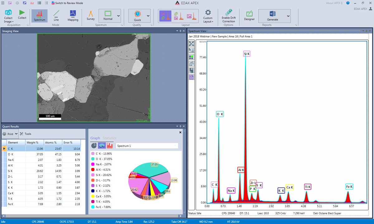
The TEAM™ Software Suite, coupled with the Octane Elect and Octane Elite Energy Dispersive Spectroscopy (EDS) Systems is the most intuitive and easy to use analytical tool available for the SEM. The workflow functions are automated by integrating years of EDAX knowledge and expertise to work for you. Startup, Analysis, and Reporting are easy because the TEAM™ software automates each task. It is the only EDS technology that combines smart decision making and guidance for the novice with advanced features for the experienced user. Now you have the intelligence of an EDS expert every step of the way. Smart features, exceptional results The TEAM™ Software Suite for the SEM was designed to save time and ensure accurate, reproducible results for a wide range of applications. Whether simply collecting a spectrum or performing complex phase analysis, the system and its touch screen capability make it easy to quickly get the results you want.
The software is built with a modern interface optimized for running in multicore and multiprocessor environments. Our TEAM™ Software Suite offers Smart Features for startup, analysis, and reporting not found in any other system. The core components that make up this functionality include: Startup Smart Track’s environmental panel monitors system status; reports operating conditions for the detector, stage, column, and more; plus allows access to advanced controls. Smart Acquisition automates routine tasks for ease of use and times savings.
Analysis Smart Phase Mapping provides a higher level of analysis by automatically collecting spectra and generating phase maps with elemental distribution and associated spectra. Point analysis and line scan with next-generation EXpert ID enable fast and easy measurement of individual and multiple points from selected areas.
Reporting Smart Data Review provides an innovative layout and project tree for quick review and reporting of images, maps, and spectra. Dynamic data editing is available in reports. The TEAM™ Software Suite contains the fundamental features needed to fully characterize samples of any nature and to efficiently output the data into Quick Reports. TEAM™ Enhanced Software Suite is a fully loaded software package that includes all the available features and options for collection, analysis, and reporting to provide the user with advanced methods to examine their materials and leads to the ultimate in materials insight. Is an optional 3D solution for EDS data, which performs both imaging and analysis operations within the same software package. EDAX offers simple, one click data transfer between the TEAM™ Software Suite and TEAM™ 3D IQ for the most comprehensive visual and analytical interpretation of EDS data available.
Java; Java Web; Spring; Android; Eclipse. Pour lire la version pdf du tutoriel. Telecharger java tete la premiere pdf file.
Is an automatic function that allows the user to search through a custom built spectrum library to find similar spectra. This greatly simplifies identification of unknowns by comparing them to a group of potential candidates and reduces the complexity of finding discrepancies and similarities between spectra.